Diseases of the gastrointestinal tract
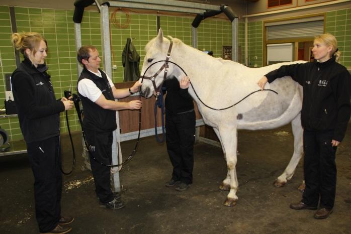
Examination of the gastrointestinal tract: The most important disease of the gastrointestinal tract in horses is colic, which can have many different causes. Colic patients make up the majority of our emergency patients. A clinical examination, blood test, rectal examination, probing of the stomach with a nasopharyngeal probe, ultrasonographic examination (Fig. 1) and abdominal cavity puncture can be used to determine the severity of the disease and make a suspected diagnosis. Simpler forms of colic such as blockages or gas build-up in the large intestine can often be treated convervatively, i.e. without surgery. Various medications, fluid therapy via nasogastric tube or infusions and exercise of the horse are available for this purpose.
If an intestinal obstruction is suspected, surgical treatment is usually unavoidable. Under anesthesia, the abdominal cavity is opened, changes in the position of the intestine are corrected and severely damaged parts of the intestine are removed. After colic surgery, the horse remains in the clinic until the stitches are removed and requires a rest period of approx. 3 months after discharge until the abdominal wall has completely healed.
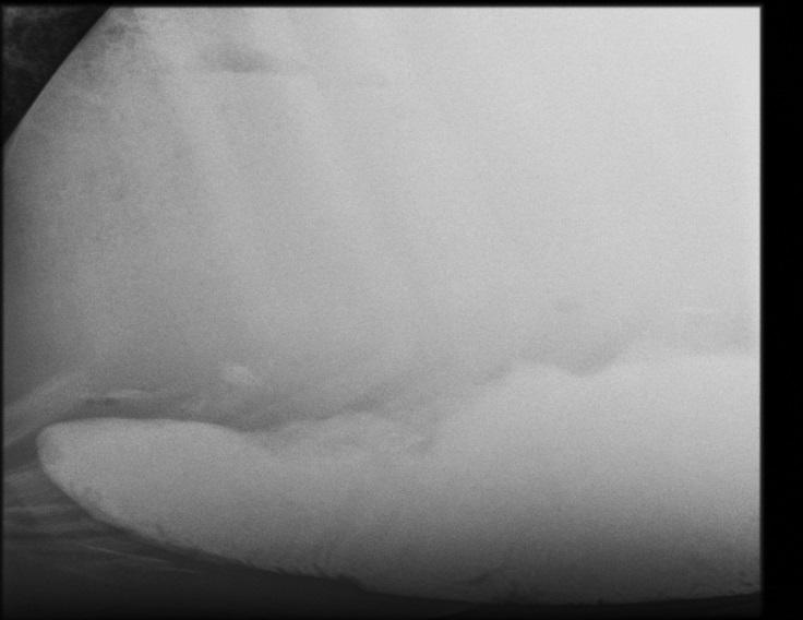
Other important diseases of the gastrointestinal tract are gastric ulcers, which are particularly common in sport horses. Symptoms include weight loss, refusal of concentrated feed, recurrent colic, increased yawning and teeth grinding. Unfortunately, the symptoms that the horse shows are not directly related to the severity of the disease. Endoscopic examination of the stomach, gastroscopy, offers the possibility of examining the stomach for gastric ulcers after a period of starvation. A very long endoscope is required for this (Fig. 2)
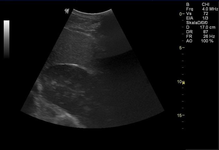
Diarrheal diseases can have various causes and in severe cases can be life-threatening. It is important to first rule out infectious causes by means of a clinical and microbiological examination, as these are infectious for other horses. Increased ingestion of sand, inflammation of the intestinal wall, parasites and problems with the absorption of nutrients from the intestine can also lead to diarrhea. In addition to the techniques mentioned in the colic examination, there are other examination options, e.g. larvae cultivation in parasitology, X-ray of the lower abdomen for sand (Fig. 3) or absorption tests to diagnose these rarer diseases.
The horse's esophagus and peritoneum can also become diseased. Pharyngeal congestion is mainly manifested by nasal discharge containing food and can lead to life-threatening pneumonia due to inhalation of food or injury to the esophagus. Peritonitis is usually accompanied by a disturbance of the general condition, colic and fever.