Diseases of the upper and deep respiratory tract
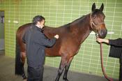
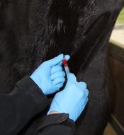
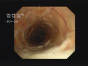
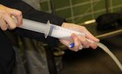
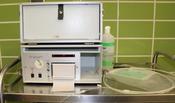
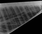
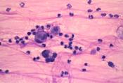
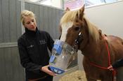
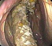
Examination of the deep respiratory tract: Diseases of the deep respiratory tract are often manifested by respiratory insufficiency, coughing or nasal discharge. The clinical examination (Fig. 1) is used to localize the location of the disease. An arterial blood gas analysis (Fig. 2) measures the oxygen content in the blood, while the endoscopic examination (Fig. 3) provides information about accumulations of secretions in the trachea (Fig. 4) and enables a sample of secretions to be taken. Furthermore, the clinic offers the possibility of bronchoalveolar lavage (Fig. 5), which is often more promising than the examination of a secretion sample from the trachea in diseases that are difficult to diagnose, as well as the measurement of interpleural pressure (Fig. 6), which provides information about the work of breathing in obstructive lung diseases. The powerful X-ray system allows shadowing of the deeper lung tissue to be visualized (Fig. 7), while inflammatory processes on the lung surface can be seen ultrasonographically. Cytologic examination of the secretions from the trachea and the bronchoalveolar lavage fluid provides information on the degree and cause of the inflammation (Fig. 8), and a microbiologic examination can also be initiated. In advanced cases, pathohistologic examination of a lung biopsy offers the possibility of assessing the prognosis for the horse. Many horses with chronic respiratory diseases can now be treated by inhalation (Fig. 9). "Lung lavage", i.e. the infusion of large quantities of fluid to mobilize viscous secretions from the airways, is also an option in severe cases of chronic obstructive bronchitis (”dampness").
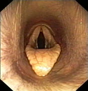
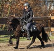
Examination of the upper respiratory tract: Diseases of the upper respiratory tract affect the nasal passages, paranasal sinuses, air sacs, larynx and pharynx. They often lead to a respiratory noise and performance insufficiency, fungal diseases of the air sacs can cause the horse to bleed to death (Fig. 10). In a standardized stress test on the lunge or under the rider, any respiratory noise is first characterized. This is followed by endoscopic examinations at rest (Fig. 11) and, if necessary, under stress (Fig. 12). Thanks to mobile systems, it is now possible to endoscope a horse under the rider or in front of the sulky while it is performing the required work during which the respiratory noise occurs. In addition to endoscopy and X-ray diagnostics, computer tomography is a modern way of precisely localizing and evaluating changes in the complex head area.
